
For more than 40 years, the clinical lab technique known as western blotting has been an important research process in all fields that relate to proteomics (the study of proteins). Preceded by the southern blot (which helped to map DNA by identifying genetic sequences using gel electrophoresis), the western blot has a proven ability to detect, count, and measure the size of targeted proteins.
The online health and wellness information hub Mobile Health Data praises the versatility of the western blot in its new overview of the western blotting technique. In addition to its well-known oncology applications, the western blot is vital to clinical research in a broad spectrum of scientific and medical disciplines. And researchers continue to discover innovative new uses for the highly adaptable western blot.
In many translational and clinical studies, western blot assays are designed, validated, and documented in collaboration with a specialized clinical trial services, helping ensure robust methodology, regulatory compliance, and reproducible results.
The western blotting process begins with the preparation of the sample to be studied. These samples can come from whole, in-tact tissue as well as tissue culture extracts. In order to preserve samples and prevent protein degradation prior to study, researchers may choose to freeze them for storage with liquid nitrogen.
Other western blot preparation methods include using a homogenizer or a lysis buffer to collect and breakdown (lyse) tissue cells. Employing a range of salts, detergents, and buffers, the lysis process can significantly improve both protein extraction and protein recognition operations.
The next key step in the western blotting process is gel electrophoresis. Gel electrophoresis allows researchers to separate proteins according to size. It is a necessary process if researchers want to measure the molecular weight, isoelectric point, and/or electric charge of each protein under study.
After chemically treating proteins on a polyacrylamide gel, researchers apply electrical current to the gel, which causes different proteins to move at different rates and ultimately separate into different bands or lanes. When the proteins have separated, researchers can place them within various lanes to determine molecular weight and antibody immunity.
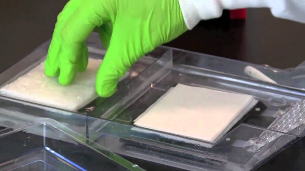
When electrophoresis is complete, the proteins on the gel are ready for transfer to a blotting membrane. This is how the western blot got its name. Blotting membrane is most commonly composed of nitrocellulose (nitric acid esters), but it can also be made from activated paper, activated nylon, and polyvinylidene difluoride.
The most common method of transferring proteins from gel to membrane is called electroblotting. This involves applying a specific voltage that adheres proteins to the membrane through the force of electromagnetism. By applying blocking buffers that range from serum albumin to nonfat dry milk, researchers can mask nonspecific potential binding sites on the membrane to eliminate false antibody positives and ensure unambiguous results.
Researchers can bind a secondary antibody to the proteins under study that reveals protein quantity using radioactive, colorimetric, fluorescent, or (most commonly) chemiluminescent means. Chemiluminescent agents produce a luminescence that researchers can use to determine overall protein quantity. Using a light source and a charged-couple device camera, researchers can measure this luminesce on photographic film.
After researchers create an image from a western blot, they can analyze that image according to optical density (densitometry) to measure the relative quantity of a protein against a predetermined control quantity and/or the quantity of other protein samples. When juxtaposing different samples, researchers should ensure that they have all been run on the same blot. This is due to the fact that significant disparities can occur among different blots.
To analyze various bands according to optical density, researchers can use the imaging software that came with their charged-couple device cameras or acquire other specialized software programs. This software allows researchers to either manually or automatically analyze bands to determine relative levels of protein expression and concentration.